Journal of Medical Imaging

Editor-in-Chief: Bennett A. Landman, Vanderbilt University, USA

The Journal of Medical Imaging (JMI) allows for the peer-reviewed communication and archiving of fundamental and translational research, as well as applications, focused on medical imaging, a field that continues to benefit from technological improvements and yield biomedical advancements in the early detection, diagnostics, and therapy of disease as well as in the understanding of normal conditions.

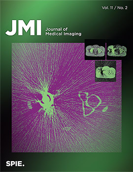
On the cover: "Dual-energy computed tomography imaging with megavoltage and kilovoltage X-ray spectra" by Giavanna Jadick, Geneva Schlafly, and Patrick J. La Rivière, in Volume 11 Issue 2.

The Journal of Medical Imaging (JMI) allows for the peer-reviewed communication and archiving of fundamental and translational research, as well as applications, focused on medical imaging, a field that continues to benefit from technological improvements and yield biomedical advancements in the early detection, diagnostics, and therapy of disease as well as in the understanding of normal conditions.  The scope of JMI includes in-depth studies of: 

    • Imaging physics and acquisition system advances across various modalities and spatial scales such as x-ray radiography, CT (computed tomography), ultrasonic imaging, nuclear medicine imaging including PET (positron emission tomography) and SPECT (single photon emission computed tomography), MRI (magnetic resonance imaging), optical imaging including OCT (optical coherence tomography), photoacoustic imaging, and combined acquisition systems such as PET/CT and PET/MRI
    • Tomographic reconstruction algorithms (such as those in CT, MRI, and OCT)
    • Advances in the customization and application of image processing and deep learning along various elements of the imaging chain, such as detector design, tomographic reconstruction, artificial intelligence, and workflow
    • Advances in computer-aided diagnosis and quantitative image analysis including customization and application of computer vision, machine learning, and deep learning in decision support
    • Advances in visualization and modeling of the human eye-brain system, including studies in image perception and observer performance
    • Advances in technology assessment methodology and applications
    • Advances in Picture Archiving and Communications Systems (PACS)
    • Advances in ultrasonic imaging including sonography and photoacoustic imaging
    • Advances in image-guided systems and targeted procedures, e.g., in surgical planning, radiation treatment planning, and theranostics
    • Advances in digital and computational pathology, from acquisition of pathology data to its management, analysis, and interpretation by observers; specific topics include image acquisition, display, interpretation, computer-aided diagnosis, quantitative image analysis of pathology specimens, and the interaction between machine learning systems and human intelligence in the analysis of microscopic images
    • Advances in biomedical applications of all imaging modalities to further understanding of disease mechanisms as well as normal physiology

    Details

      About the Journal

      ISSN: 2329-4302
      E-ISSN: 2329-4310
      Publisher: SPIE
      Frequency: Article-at-a-time publication. Bi-monthly, since 2020 (6 issues/year)
      Format: Online
      Year established: 2014

      Metrics

      CiteScoreTM 2022: 5.7
      Impact Factor:* 2.4

        * Source: Clarivate Analytics, Journal Citation Reports (2023)


      Abstracting and Indexing

      • PubMed
      • PubMed Central
      • Web of Science (SCIE)
      • Inspec
      • Scopus
      • Ei Compendex
      • Embase

      Editorial Office

      SPIE
      P.O. Box 10
      Bellingham, WA 98227-0010 USA
      Tel: +1 360 676 3290
      Fax: +1 360 647 1445
      Email: journals@spie.org


    Back to Top