|
During the last few decades of the twentieth century, partly in concert with the increasing availability of relatively inexpensive computational resources, medical imaging technology, which had for nearly 80 years been almost exclusively concerned with conventional film/screen x-ray imaging, experienced the development and commercialization of a plethora of new imaging technologies. Computed tomography, MRI imaging, digital subtraction angiography, Doppler ultrasound imaging, and various imaging techniques based on nuclear emission (PET, SPECT, etc.) have all been valuable additions to the radiologist's arsenal of imaging tools toward ever more reliable detection and diagnosis of disease. More recently, conventional x-ray imaging technology itself is being challenged by the emerging possibilities offered by flat panel x-ray detectors. In addition to the concurrent development of rapid and relatively inexpensive computational resources, this era of rapid change owes much of its success to an improved understanding of the information theoretic principles on which the development and maturation of these new technologies is based. A further important corollary of these developments in medical imaging technology has been the relatively rapid development and deployment of methods for archiving and transmitting digital images. Much of this engineering development continues to make use of the ongoing revolution in rapid communications technology offered by increasing bandwidth.
A little more than 100 years after the discovery of x rays, this three-volume
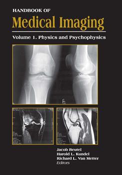
Handbook of Medical Imaging is intended to provide a comprehensive overview of the theory and current practice of Medical Imaging as we enter the twenty-first century. Volume 1, which concerns the physics and the psychophysics of medical imaging, begins with a fundamental description of x-ray imaging physics and progresses to a review of linear systems theory and its application to an understanding of signal and noise propagation in such systems. The subsequent chapters concern the physics of the important individual imaging modalities currently in use: ultrasound, CT, MRI, the recently emerging technology of flat-panel x-ray detectors and, in particular, their application to mammography. The second half of this volume, which covers topics in psychophysics, describes the current understanding of the relationship between image quality metrics and visual perception of the diagnostic information carried by medical images. In addition, various models of perception in the presence of noise or "unwanted" signal are described. Lastly, the statistical methods used in determining the efficacy of medical imaging tasks, and ROC analysis and its variants, are discussed.
Volume 2, which concerns Medical Image Processing and Image Analysis, provides descriptions of the methods currently being used or developed for enhancing the visual perception of digital medical images obtained by a wide variety of imaging modalities and for image analysis as a possible aid to detection and diagnosis. Image analysis may be of particular significance in future developments, since, aside from the inherent efficiencies of digital imaging, the possibility of performing analytic computation on digital information offers exciting prospects for improved detection and diagnostic accuracy.
Lastly, Volume 3 describes the concurrent engineering developments that in some instances have actually enabled further developments in digital diagnostic imaging. Among the latter, the ongoing development of bright, high-resolution monitors for viewing high-resolution digital radiographs, particularly for mammography, stands out. Other efforts in this field offer exciting, previously inconceivable possibilities, e.g., the use of 3D (virtual reality) visualization for surgical planning and for image-guided surgery. Another important area of ongoing research in this field involves image compression, which in concert with increasing bandwidth enables rapid image communication and increases storage efficiency. The latter will be particularly important with the expected increase in the acceptance of digital radiography as a replacement for conventional film/screen imaging, which is expected to generate data volumes far in excess of currently available capacity. The second half of this volume describes current developments in Picture Archiving and Communications System (PACS) technology, with particular emphasis on integration of the new and emerging imaging technologies into the hospital environment and the provision of means for rapid retrieval and transmission of imaging data. Developments in rapid transmission are of particular importance since they will enable access via telemedicine to remote or underdeveloped areas.
As evidenced by the variety of the research described in these volumes, medical imaging is still undergoing very rapid change. The editors hope that this publication will provide at least some of the information required by students, researchers, and practitioners in this exciting field to make their own contributions to its ever-increasing usefulness.
|


